Of all the branches of science, my absolute favorite is that of neurology! In my opinion, hands down, it is the most interesting part of the human body! I’m sure you can imagine my delight when it came time for this field of study in my Anatomy and Physiology class. For clarification purposes, I resembled a small child…in a toy store…after winning the lotto. Yep- I was that excited! This blog post is going to be about the Nervous System in a nutshell. In actuality, I could create a blog site all by itself that is centered on the nervous system, add a new post every week, and be able to keep it updated for a super long period of time. But, for your sake (as I’m sure not all of you share the same passion for this intricate body system as I do) I will keep it down to as basic as I can. Neurology in a nutshell is going to explain what comprises the Nervous System, how it works, and why it’s important. I’m so excited, so let’s begin!
Most of you who are routine visitors to my blog will know that I created a video on the Neuromuscular Junction a little while back. I suggest you go watch the video, with link here, as it visually explains how neurons work (which is what we’re going to talk about momentarily). Many of the terms that I will be discussing in this post are in the video (besides, videos are always better than paragraphs, right?). It is a wonderful supplement that I highly recommend you watch to enrich your understanding of the Nervous System. Check it out!
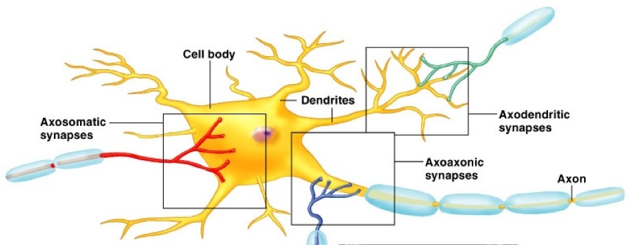
First off, the Nervous System is divided into two main sections: The Central Nervous System (CNS), and the Peripheral Nervous System (PNS). The CNS is comprised of the brain and spinal cord. The PNS is all of the nerves that branch out from the CNS, such as the nerves in your limbs and organs. Regardless of which section you are in, there are nerve cells, which are called neurons. The neuron is modeled below:

A neuron, as you can see, is comprised of a cell body, tentacle-like dendrites, and synapses. The cell body contains the nucleus of the neuron cell. The dendrites are responsible for connecting to other neurons. The synapses, which we will go more in-depth on in a minute, are how the neurons communicate. But before we go into that step, I want to point out the two main types of synapses. There are axondendritic synapses, which is when a neuron connects to the dendrites of a different neuron. There are also axosomatic synapses, where the axon of a neuron attaches directly to the neuron body itself (called a soma). Being the most direct, the latter is more powerful of a signal. Other types of connections include axoaxonic – connection between two axons, dendrodendritic – connection between two dendrites, and dendrosomatic – connection between dendrites and somas. These are important to understand as it demonstrates the massive connectivity, and large interwoven network, the nervous system has created. (Take notice of the axosomatic, axoaxonic, and axodendritic synapses in the above photo).
Now I have a confession to make. You know how I just finished explaining how the neurons “connect” with each other? Well, in all reality, they never actually touch each other. Synapses are the junctions where neurons “meet up” and communicate, but do so without physical contact. These synapses, known as chemical synapses, involve a lengthy process of releasing chemicals into a fluid-filled space between neurons known as the synaptic cleft. Maybe a conceptual picture (or a glance at my video!) will help:
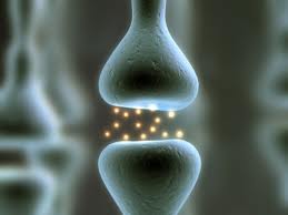
You can see how the neurons do not touch with each other. Instead they release chemicals that travel across the synaptic cleft. (On a side note, there is a type of synapse in the body called an electrical synapse. These neurons DO connect with each other. This connection does not have a gap like in the picture above. However, they are very rare, as each neuron has to fire if the previous one does. Their function wake you up, provide mental attention, provide emotion, and benefit memory). No matter which type of synapse you refer to, there are certain terms for the cellsas they deliver messages. A cell that has a “message”, called an impulse, traveling toward the synapse is called a presynaptic neuron. The neuron receiving the impulse from a previous neuron is called a postsynaptic neuron. As impulses travel in a linear fashion, it is easy to see how one end of a neuron is presynaptic, while the other is postsynaptic. Make sense?
Ok, so a quick recap: we now know what the neurons look like, and how they connect to each other. We still need to uncover how they communicate at a synapse and with what they are communicating with. We’re going to start with their messages themselves, and then explain in great detail how the messages travel.
These messages are called impulses. Impulses are determined by two concepts known as Resting Potential, and Action Potential (I am being very serious when I say that now would be a good time to switch over tothe video. There is a wonderful visual representation within it that explains these very clearly!). Resting potential is when the inside of a neuron is neutrally charged, and the outside is positively charged (this is when the neuron is “resting”; there is no major impulse happening). For your information, as this information will become relevant later, the negative charge within a neuron is -70mV. Action potentials are when there is a sudden switch in the polarity of the neuron (the inside becomes positive, the outside negative). This action potential travels down the neuron, in a sort of ripple effect (the individual charges only switch polarity when the previous one in line has done so. They then switch back after the “wave” has passed. Trust me- watch my video!). This is how messages are sent down the neurons. What is occurring chemically during these action potentials is toward the end of this post.
As the initial picture of a neuron showed, there is a myelin sheath (also known as a Schwann cell) that surrounds the axon of neurons. The purpose of this cell, which surrounds the axon in a spiraled sort of coil, is to increase the speed in which a message travels through the neuron (it can actually increase it 100 times the regular speed). You see, for these impulses to travel through a neuron, they have to undergo the action potential process I explained earlier. However, with the myelin sheath, this action potential never fires. So the impulse can pass much quicker, as it only has to fire as an action potential between myelin sheath cells (which is dramatically quicker!). Talk about efficiency!

Furthermore, there are two types of actionpotentials that can occur. One is called Excitatory Postsynaptic Potential (EPSP). This is when there is a minor increase in the positive polarity of the inside of the neuron. Basically, this is the start of an action potential. A diagram is shown below. (The “Threshold” line occurs at -55 mV. This is the point where an EPSP transforms into an actual action potential, and fires an impulse. Otherwise, without reaching the threshold, it will not fire. Think of it as a tiny tremor, if you will).

On the contrary to an EPSP is an Inhibitory Postsynaptic Potential (IPSP). The IPSP does the exact opposite of the EPSP- it makes the neuron harder to fire by increasing the negative polarity within the cell. The farther from the threshold, the more energy it takes to fire an impulse.

You are doing a fantastic job by making it this far! I know, this is a lot of information. Correction: this is a TON of information (if you couldn’t tell, I lied about keeping it short, such as in a “nutshell”, as the title states. Sorry!). But trust me, reading this will make you the smartest person on your block! Hang in there! So now that we have knowledge into the different types of potentials, and how they correlate to impulses, we have to add two more components to the mix: when and where the EPSP and IPSP’s occur (what time they occur, and how quickly in proximity to eachother they occur, respectively). This is crucial to determining whether the polarity of the neurons will reach the Threshold, and be able to fire an impulse. Consider the four charts below, labeled a, b, c, and d.

The first chart, “a”, has two EPSP pulses coming from a single axon terminal. However, they are unsynchronized, and are far apart enough that the threshold limit was not reached. The neuron, as a result, won’t fire. Chart labeled “b” has two EPSP pulses from the same terminal that fire in quick succession to each other (known as temporal summation), resulting in the polarity tocross the threshold line and fire the neuron. Chart “c” demonstrates what is known as spatial summation. This is when multiple EPSP pulses from multiple neuron terminals fire in unison to allow the polarity to cross the threshold, and achieve an impulse fire. Chart “d” demonstrates how an EPSP and an IPSP pulse fire in close secession, resulting in no change and no impulse (as the positive charged EPSP cancels with the negative charged IPSP). So as you can see, the when and where are vital to determining whethera neuron will fire an impulse.
We now have all the essential puzzle pieces but one: how do synapses, and by extension neurons, communicate with each other? So far we have said that impulses travel between synapses to pass along messages. While thisis true, there is so much more involved than just that. Sometimes, however, a picture speaks a thousand words. Take a look:

This is a visual representation of how the neurons communicate with each other. You can see the axon terminal of the presynaptic neuron, the synaptic cleft, and the cell membrane of the postsynaptic membrane. To start, the impulse travels down the nerve axon into the axon terminal. This impulse releases calcium, which then releases neurotransmitters into the synaptic cleft (neurotransmitters are the chemicals that transfer the signal. In cases of muscles, for example, the neurotransmitter Acyetylcholine is released). The neurotransmitter bonds with ion channels, which then are opened, and allow sodium to enter the neuron. My video, once again, hasa great description of this seems confusing to you!
Let’s look at the ion channel more closely (a picture follows). The ion channel is closed initially, ensuring nothing can enter the neuron. However, the presence of a a neurotransmitter such as Acytylcholine causes it to open and allow ions to enter the neuron. There is then an enzyme, which using our example would be Acetylcholinesterase, that breaks apart the neurotransmitter and closes the channel. There are two important things to understand about these ion channels. The first is that there is a concept called tetanus, which is when the muscles lock up and contract without release. This is caused by the neurotransmitter remaining in the ion channel receptor, which causes the cell to continue firing. The other important concept is that these ion channels are thought to be a component of musclememory and learning. Let’s take playing the drums as an example. When you first start, you don’t play very well, as your body isn’t used to using its muscles in that way. At this point, there are very few ion channels. But, as you keep practicing, the number of ion channels increase (allowing quicker receptiveness), which leads to “muscle memory”. The more channels to recept the neurotransmitter, the more quickly and fluently the action will occur. Pretty neat, huh?

The last thing I’m going to mention about the synapses is that there is a higher concentration of chemicals sodium and calcium on the outside of a neuron, and a higher concentration of potassium on the inside of neurons. This is what causes the differences in charges that I described earlier. However, along with the ion channels mentioned above, there are also pumps that use ATP energy to move sodium, calcium, and potassium in and out of the cell (the moving of these substances involve differences in gradients, and the natural urge to diffuse into areas with less concentration). The moving of these substances are what cause the nerve impulse in the first place (basically, this is the explanation as to why the charges in action potentials switch during an impulse). I waited until the end to bring it up because it is crucial to understanding how the synapses of neurons work.
Congratulations! This marks the end of my blog post on the Nervous System. You should feel proud for reaching this point! We have covered a lot in this post: what the nervous system is, what comprises it, howit communicates and by what means it communicates with! And that is without mentioning the plethora of technical terminology or detours we covered either! All in all, I hope that you have gained a new appreciation for the sophistication, and inter-woven complexity, of the Nervous System. It truly is the system responsible for keeping the entire rest of the body operating! Now do you see why I’m so entranced by the unique anatomy and physiology of this body system? Everything you do, or ever think about doing, is possible because of the flawless workings of your Nervous System!