Plant materials and microorganisms
T. bellirica dried fruit samples were procured from a traditional medicine store in Kandy, Sri Lanka and authenticated by Mrs. N.P.T. Gunawardena, a plant taxonomist at National Herbarium at Peradeniya Royal Botanical Garden, Sri Lanka. A voucher specimen (# 816) has been deposited in the National Herbarium at Peradeniya Royal Botanical Garden. The bacterial strains, S. aureus ATCC 25923 and NCTC 6571, E. coli ATCC 25922 and extended spectrum β-lactamase (ESBL)-producing K. pneumoniae ATCC 700603 and MDR bacteria were obtained from the archives of the Department of Microbiology, Faculty of Medicine, University of Peradeniya. The MDR bacterial strains have been isolated from clinical samples and saved in the archives of the Department of Microbiology as a part of routine practice. Since the isolates are used as a source of bacterial strains only, the isolates stay anonymous and the records are not associated with specific pathology or specific individuals. The bacterial strains were handled following the standard health/safety procedures. All bacterial strains were stored at − 81 °C (Thermo Scientific, USA). Disc diffusion antibacterial sensitivity testing was carried out on 35 bacterial isolates according to the Clinical and Laboratory Standard Institute method [25] with antibiotics representing different classes of antibiotics: penicillins (ampicillin), cephalosporins (cefuroxime, cefotaxime, ceftazidime, cefipime), carbapenems (imipenem, meropenem), quinolones (ciprofloxacin), monobactams (aztreonam) and aminoglycosides (amikacin, netilmicin, gentamicin). The following MDR bacterial strains were used for screening aqueous and organic extracts of T. bellirica fruit for antibacterial activity: 8 strains of MRSA with minimum inhibitory concentration (MIC) of oxacillin ≥128 mg/L and 2 strains each of ESBL-producing E. coli, MDR Acinetobacter spp., MDR K. pneumoniae and MDR P. aeruginosa.
Preparation of extracts
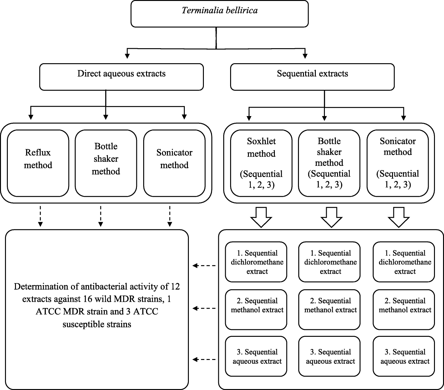
The fruits of T. bellirica were further dried in the laboratory at room temperature for 1 week and ground using an electric grinder to obtain powdered plant material. The plant extracts were prepared from the powdered plant material to obtain 3 direct aqueous extracts—using reflux, bottle shaker and sonicator methods—and 9 sequential extracts using Soxhlet, bottle shaker and sonicator methods (Fig. 1). Each sequential extraction procedure involved three solvents (dichloromethane, methanol and water) of increasing polarity. In each extraction procedure—direct aqueous or sequential—the ratio of dried fruit (weight in g) and extracting solvent (volume in mL) was maintained as 1:10 [26]. The procedures employed for the preparation of direct aqueous extracts and sequential extracts are given below.

An overview of different methods used for extracting dried pericarp of Terminalia bellirica fruit
Direct aqueous extracts
Aqueous extracts were obtained from the powdered plant material using the following three methods: an aliquot of the powdered material (40 g) was suspended in distilled water (400 mL) and heated under reflux for 6 h; a second aliquot of the powdered material (40 g) was also extracted into distilled water (400 mL) under ambient temperature using a bottle-shaker (GFL 3016, Germany) for 24 h; a third aqueous extract was obtained by ultrasound sonication (Branson 2510, USA) of the powdered material (40 g) in distilled water (400 mL) at ambient temperature for 3 h. Each aqueous extract was centrifuged, and the supernatant freeze-dried to obtain a light brown powder.
Sequential extracts
The powdered plant material (40 g) was packed in a cellulose thimble, placed in the extraction tube of a Soxhlet apparatus and extracted with dichloromethane (400 mL) for 6 h, and then the extraction was continued with methanol (400 mL) for a further 6 h. The plant residue was dried and heated in distilled water (400 mL) under reflux for 6 h. Another aliquot of powdered plant material (40 g) was also extracted into dichloromethane (400 mL) followed by methanol (400 mL) and distilled water (400 mL) at ambient temperature for 24 h using a bottle-shaker. A third set of sequential extracts was obtained by ultrasound sonication of the powdered material (40 g) in dichloromethane (400 mL) followed by methanol (400 mL) and distilled water (400 mL) at ambient temperature for 3 h using an ultrasound sonicator. The solvent of each dichloromethane and methanol extract was removed under reduced pressure using a rotary evaporator (Heidolph, Laborota 4000, Germany). Each sequentially obtained aqueous extract was centrifuged and the supernatant freeze-dried to obtain a crude powder. The powder obtained from each extract was checked for sterility on a nutrient agar plate and stored in air tight universal bottles at − 20 °C till further testing. A schematic diagram of the extraction procedures is given in Fig. 1.
Screening for antibacterial activity
The aqueous and organic solvent extracts of T. bellirica fruits were screened for antibacterial activity using the cut-well diffusion method [26]. Briefly, bacterial suspensions of test and control organisms were adjusted to McFarland turbidity of 0.5 (approximately 1 × 10⁸ cfu/mL) and inoculated onto Mueller Hinton agar (MHA, Oxoid, Hampshire, England). The plates were left at room temperature for 30 min after which 12-mm diameter wells were bored in the agar and the bottom sealed with molten MHA. The organic extracts were dissolved with the aid of 10% (v/v) aqueous dimethyl sulfoxide (DMSO, BDH, England). Using a template, aliquots of each reconstituted extract (10 mg/mL) were pipetted into the wells and the plates incubated aerobically at 35 °C for 24 h. The diameter of the zone of inhibition (ZOI) around the well was measured along with the well. Each screening was carried out in triplicate and the mean diameter of the ZOI was recorded.
Minimum inhibitory concentration (MIC)
The MICs of the aqueous and organic fruit extracts were determined by the agar dilution method [27]. Briefly, stock solutions of concentration 20 and 10 mg/mL were prepared from each extract and diluted with molten MHA (45 °C) to obtain a series of concentrations 5, 4, 2, 1, 0.5, 0.25 and 0.125 mg/mL, which were poured into sterile petri dishes and allowed to set. A 2-μL drop of each test and control organism prepared as stated above was inoculated onto each plate using a template. The plates were read after incubation at 35 °C for 24 h. The lowest concentration of extract that exhibited no visible growth was recorded as the MIC for each organism.
Determination of DPPH free radical scavenging activity
DPPH radical (Sigma Aldrich, USA) scavenging activity was determined following a procedure described by Zhang et al. [28], with slight modifications. Briefly, 100 μL of each extract at various dilutions (50–3 ppm) was mixed with 100 μL of 1.6 mM DPPH solution in flat-bottom 96-well microtiter plates. The mixture was shaken for 1 min, kept for 30 min in the dark and the absorbance measured at 517 nm in an automated microplate reader (Biochrome UVM 340-Elisa Reader, USA). All determinations were performed in triplicate. L-ascorbic acid was used as a positive control.
The percentage scavenging effect was calculated as: % Scavenging rate = [{A0 − (A1 − A2)} / A0] × 100%, where A0 is the absorbance of the control (without sample) and A1 is the absorbance of sample in the presence of the DPPH, A2 is the absorbance of sample without DPPH radical (blank absorbance). The scavenging ability of the samples was expressed as EC50 value, the effective concentration at which 50% of DPPH radicals were scavenged; the EC50 value was calculated from the curve of percentage scavenging activity (%) versus concentration of the respective sample.
Determination of total phenolic content (TPC)
TPC of solvent extracts of T. bellirica was determined using Folin-Ciocalteu reagent following a procedure described by Antolovich et al. [29], with minor modifications. Briefly, 20 μL of each extract was mixed with 100 μL of 1:10 Folin-Ciocalteu’s reagent (Merck, Germany) followed by the addition of aqueous Na2CO3 (80 μL, 7.5%). The assay was carried out in the automated microplate reader. After incubation at room temperature for 1 h in the dark, the absorbance at 765 nm was recorded. A standard curve for gallic acid solution (1, 2, 3, 4, 5, 6, 7, 8, 9, 10 and 20 ppm) was prepared using the same procedure. TPC was expressed as mg gallic acid equivalents per gram of dried extract (mg GAE/g).
Cytotoxic assay of the most potent antibacterial extract of T. bellirica
Of the 12 extracts obtained from T. bellirica fruits, the direct aqueous extract (reflux method) was identified as the extract having the highest antibacterial potency. The cytotoxicity of this extract was evaluated using baby hamster kidney (BHK-21) cells available from the Animal Virus Laboratory, Veterinary Research Institute, Polgolla, Sri Lanka. The cytotoxic assay was performed by the method described by Jirasripongpun et al. [30], with minor modifications. Briefly, the BHK-21 cells (1 × 10⁵ cells/mL) were seeded onto a 6-well plate (Falcon, New Jersey, USA) containing Minimum Essential Medium Eagle (MEM) (Sigma-Aldrich, USA) supplemented with 10% fetal calf serum (Sigma Aldrich, USA) to provide confluence after 10–12 h incubation. The spent medium was removed, and the volume adjusted to 10 mL with new medium (MEM) containing a solution of the direct aqueous extract (reflux method) of T. bellirica such that the concentration of the extract was 4, 2, 1, 0.5, 0.25 and 0.125 mg/mL at separate runs. Distilled water and 20% aqueous DMSO (Sigma Aldrich, USA) containing plates served as negative and positive controls, respectively. The cultures were further incubated for 48 h and samples were counted for cell viability each day using Tryphan Blue exclusion method and hemocytometer. Each experiment was carried out in triplicate and averaged percent cell viability was plotted against concentration of T. bellirica aqueous extract. The 50% inhibition concentration (IC50) reflects the concentration of T. bellirica extract causing a 50% decrease in cell viability.
Statistical analysis
All the experiments were carried out in triplicate. The data is expressed as mean ± standard deviation (SD). To determine the significant differences between values, analysis of variance (ANOVA) and Duncan’s multiple range tests were performed. Significance of difference was defined at 5% level (p http://systasoftware.com) and R-3.2.0 (R Software Inc. Vienna, Austria, http://www.r-project.org). The correlation analysis was performed between antioxidant activity (mean EC50) and total phenolic content.